>
产品中心 >
Functional_antibody >
absoluteantibody/Anti-FceR1 alpha [Mar-1]/200 μg/Ab01187-23.0
![absoluteantibody/Anti-FceR1 alpha [Mar-1]/200 μg/Ab01187-23.0](https://www.ebiomall.cn/images/no_picture.gif)
UniProt Accession Number of Target Protein: P20489
Alternative Name(s) of Target: Fc epsilon RI alpha; FceRIa; FceRI alpha; FceRI-α; high affinity IgE receptor; High affinity immunoglobulin epsilon receptor subunit alpha; Fcer1a; Fce1a; Fc-epsilon RI-alpha; FcERI; IgE Fc receptor subunit alpha
Specificity: This antibody specifically targets the non-signalling alpha chain of the FceR1 complex.
Application Notes: This antibody has been used in flow cytometric analysis of FceR1 alpha cell surface expression by basophils (Min et al, 2004; Chen et al, 2009), and immunohistochemical analysis of epidermal sheets (Holzmann et al, 2004). It has also been used in immunoprecipitation analysis of cell lysates from lung cDCs and MC/9 mast cells (Grayson et al, 2007). In vitro incubation of basophils with this antibody blocks subsequent MAR-1 staining, but does not activate basophil IL-4 production (Sokol et al, 2008). Intravenous treatment of mice with this antibody results in basophil depletion in peripheral blood, spleen, bone marrow and liver, but not skin or intraperitoneal mast cell depletion (Sokol et al, 2008). Following treatment with this antibody, basophil-depleted mice display impaired Th2 differentiation upon papain immunisation (Sokol et al, 2008). However, MAR-1 treated mice respond normally to high-dose intravenous Ig in both the K/BxN serum transfer arthritis and collagen-induced arthritis models, despite basophil depletion (Campbell et al, 2014).
Antibody first published in:Holzmann et al.A model system using tape stripping for characterization of Langerhans cell-precursors in vivo.J Invest Dermatol. 2004 May;122(5):1165-74.PMID:15140219Note on publication:Describes the original use of this antibody in immunohistochemical analysis of epidermal sheets.


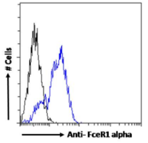
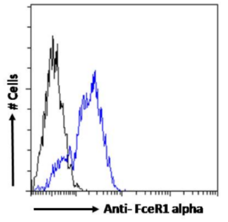
Flow-cytometry using the anti-FceR1 alpha antibody Mar-1 (Ab01187) Mouse splenocytes were stained with anti-Fluorescein IgG antibody (4-4-20; isotype control, black line) or the rabbit IgG-chimeric version of Mar-1 (Ab01187-23.0, blue line) at a dilution of 1:100 for 1h at RT. After washing, bound antibody was detected using a goat anti-rabbit IgG AlexaFluor® 488 antibody at a dilution of 1:1000 and cells analyzed using a FACSCanto flow-cytometer.


